Saturday, 16 October 2021

Actuarial (versus actual)

«Its use is often extended (...) to include the Kaplan-Meier (KM) method, (...). Actual is a new term (...) to refer to a technique with several designations in the statistical literature, including cumulative incidence and crude, unadjusted, absolute, or observable probability [1].»
«Actuarial analysis, (...), is used to describe and compare survival probabilities by allowing for partial survival times (censoring). (...) The actuarial event-free (survival) curve for a nonfatal event, such as structural valve deterioration (SVD) of porcine valves, estimates the event-free probability for a population in which death has been eliminated. This overestimates the percentage of valves that will actually fail, because many patients die before the valve fails [2].»
«Unlike cumulative incidence [actual estimates], the KM attempts to predict what the latent failure probability would be if death were eliminated. To do this, the KM method assumes that the risk of dying and the risk of failure are independent. But this assumption is not true for many cardiac applications in which the risks of failure and death are negatively correlated (ie, patients with a higher risk of dying have a lower risk of failure, and patients with a lower risk of death have a higher risk of failure, which is a condition called informative censoring) [3].»
«When used for nonfatal events such as SVD, actual analysis estimates the percentage of patients who will have SVD (or the probability that an individual patient will experience SVD). The KM (actuarial) method attempts to estimate the percentage of SVD that would occur if patients never died. Also, KM depends on the assumption that death and SVD are independent, which they are probably not [1].»

Bibliographic references:
[1] Grunkemeier GL, Wu Y. Actual versus actuarial event-free percentages. Ann Thorac Surg. 2001 Sep;72(3):677-8. Available at: https://doi.org/10.1016/s0003-4975(01)03059-4.
[2] Grunkemeier GL, Jamieson WR, Miller DC, Starr A. Actuarial versus actual risk of porcine structural valve deterioration. J Thorac Cardiovasc Surg. 1994 Oct;108(4):709-18. PMID: 7934107. Available at: https://doi.org/10.1016/S0022-5223(94)70298-5.
[3] Grunkemeier GL, Jin R, Eijkemans MJ, Takkenberg JJ. Actual and actuarial probabilities of competing risks: apples and lemons. Ann Thorac Surg. 2007 May;83(5):1586-92. Available at: https://doi.org/10.1016/j.athoracsur.2006.11.044.

Are you satisfied with the level of diversity, equity and inclusion in your workplace?


If you have not done so yet, we invite you to take this survey and let us know what challenges you have been facing in your career development.

Your contribution will help us get a detailed and global picture of how the oncology workforce is doing when it comes to issues related to diversity, equity, and inclusion at work.

The survey is anonymous and should take less than 15 minutes to complete. Findings will be presented at ESMO 2022.

Take the survey →

Thank you.

ESMO Women for Oncology

Sunday, 3 October 2021

QUANTEC (quantitative analysis of normal tissue effects in the clinic)

It summarizes the currently available three-dimensional dose/volume/outcome data to update and refine the normal tissue dose/volume tolerance guidelines provided by the classic "Emami" paper [1,2].

Bibliographic references:
[1] Marks LB, et al. Use of normal tissue complication probability models in the clinic. Int J Radiat Oncol Biol Phys. 2010 Mar 1;76(3 Suppl):S10-9. Available at: https://doi.org/10.1016/j.ijrobp.2009.07.1754.
[2] Emami B, et al. Tolerance of normal tissue to therapeutic irradiation. Int J Radiat Oncol Biol Phys. 1991 May 15;21(1):109-22. Available at: https://doi.org/10.1016/0360-3016(91)90171-y.

NTCP (normal tissue complication probability)

Rare cancers

The definition of rare tumors is not consensual. Rare cancers are defined as an incidence of fewer than 6 cases per 100.000 individuals per year (RARECARE workin group, Gatta et al. Eur J Cancer 2011).

According to DeSantis et al. ACS Atlanta. CA Cancer J Clin 2017, overall, approximately 20% of patients with cancer in the United States are diagnosed with a rare cancer. Based on the RARECARE workin group, Gatta et al. Eur J Cancer 2011, rare cancers are about 22% of all cancer diagnoses in Europe and 24% of the total cancer prevalence.

Rare cancers include > 300 histological subtypes and may affect all organs (Morfouace et al. EORTC, ESMO Open 2020). In addition, they can be grouped within 12 families of rare cancers (Casali et al. Fondazione IRCCS INT, Milan, ESMO Open 2020):

  1. Epithelial tumours of head and neck:
    • Larynx;
    • Hypopharynx;
    • Nasal cavity and sinuses;
    • Nasopharynx;
    • Major salivary glands and salivary gland type tumours;
    • Oropharynx;
    • Oral cavity and lip;
    • Eye and adnexa;
    • Middle ear.
  2. Epithelial digestive tumours:
    • Small intestine;
    • Anal canal;
    • Gallbladder and extrahepatic biliary duct.
  3. Thoracic tumours:
    • Epithelial tumours of the trachea;
    • Thymomas and thymic carcinomas;
    • Malignant mesothelioma.
  4. Female genital tumours:
    • Non-epithelial tumours of the ovary;
    • Epithelial tumours of the vulva and vagina;
    • Trophoblastic tumours of the placenta.
  5. Male genital and urogenital tumours:
    • Tumours of the testis and paratestis;
    • Epithelial tumours of penis;
    • Extragonadal germ cell tumours;
    • Epithelial tumours of renal pelvis, ureter, and urethra.
  6. Skin cancers and non-cutaneous melanoma:
    • Mucosal melanoma;
    • Uveal melanoma;
    • Adnexal skin carcinomas;
    • Kaposi sarcoma.
  7. Sarcomas:
    • Soft tissue sarcoma;
    • Bone sarcoma;
    • Gastrointestinal stromal tumours.
  8. Neuroendocrine tumours:
    • Gastrointestinal pancreatic;
    • Lung;
    • Other sites.
  9. Endocrine organ tumours:
    • Thyroid cancers;
    • Parathyroid cancer;
    • Adrenal cortex cancer;
    • Pituitary gland cancer.
  10. Central nervous system tumours:
    • Glial tumours and others;
    • Malignant meninioma;
    • Embryonal tumours.
  11. Paediatric tumours:
    • Hepatoblastoma;
    • Neuroblastoma and ganglioneuroblastoma;
    • Nephroblastoma;
    • Odontogenic malignant tumours;
    • Olfactory neuroblastoma;
    • Pancreatoblastoma;
    • Pleuropulmonary blastoma;
    • Retinoblastoma.
  12. Haematological malignancies:
    • Lymphoid malinancies;
    • Myelodysplasctic syndromes;
    • Myeloproliferative neoplasms (including mastocytosis);
    • Myelodysplastic/myeloproliferative neoplasms;
    • Myeloid/lymphoid neoplasms with eosinophilia and abnormalities of PDGFRA (platelet derived growth factor receptor alpha), PDGFRB (platelet derived growth factor receptor beta), or FGFR1 (fibroblast growth factor receptor 1), or with PCM1-JAK2 (pericentriolar material 1-janus kinase 2);
    • Acute myeloid leukaemia and related neoplasms.

Then, rare cancers are not so rare, and they are associated with multiple challenges including late and incorrect diagnosis, adverse outcomes, limited clinical expertise, weak evidence for best practice, and difficulties in collecting large series for research and in carrying out clinical trials (Boyd et al. BCCA, Vancouver, Lancet Oncol 2016).

Therefore, a global strategy is needed to join efforts to increase knowledge of this group of cancers.

Tuesday, 29 December 2020

Top papers for 2020 - Radiation Oncology

Read here the most important papers in Radiation Oncology this year.
Source: Top Papers for 2020 - Radiation Oncology (shared collection) [Internet]. Read.qxmd.com. 2020 [cited 29 December 2020]. Available from: https://read.qxmd.com/collection/22121?sid=adea2753-d897-4e24-bd70-dad41e492317.

Saturday, 26 December 2020

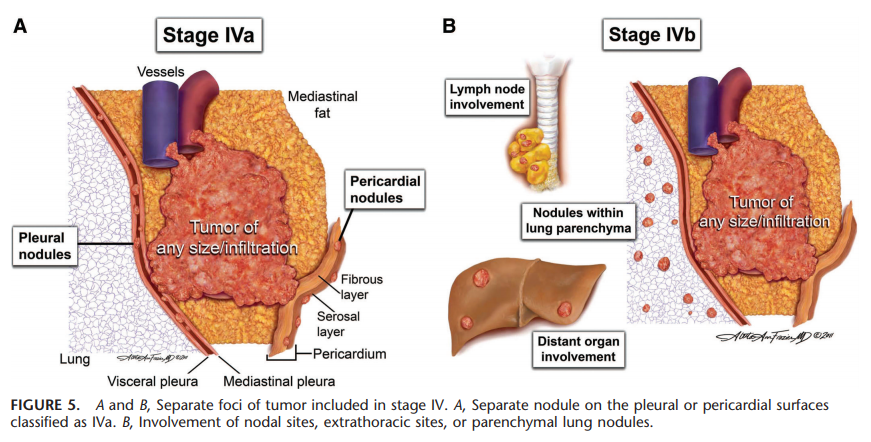
Thymoma and thymic carcinoma staging systems

Masaoka-Koga staging system of thymic tumors:

Source: Jukna A, Jasa M, Mezvevere M, et al. Inside the Mediastinum: the Morphological Spectrum and Stages of Thymic Tumours. Acta Chir Latv. 2016; 16(1):3-8. Available at: https://doi.org/10.1515/chilat-2016-0010.

Source: Detterbeck FC, Nicholson AG, Kondo K, et al. The Masaoka-Koga stage classification for thymic malignancies: clarification and definition of terms. J Thorac Oncol. 2011 Jul;6(7 Suppl 3):S1710-6. Available at: https://doi.org/10.1097/jto.0b013e31821e8cff.

The modified Masaoka staging system separates stage III in IIIa (without invasion of great vessels) and IIIb (with the invasion of great vessels).
Bibliographic reference: NCCN Clinical Practice Guidelines In Oncology (NCCN Guidelines), Thymomas and Thymic Carcinomas, Version 1.2021 - December 4, 2020 [Internet]. Nccn.org. 2020 [cited 26 December 2020]. Available from: https://www.nccn.org/professionals/physician_gls/pdf/thymic.pdf.

The 8th edition thymic TNM stage classifications:

Source: Detterbeck FC. Clinical implication of the new TNM classification of thymic malignancies. J Thorac Dis. 2018 Aug;10(Suppl 22):S2692-S2695. Available at: https://doi.org/10.21037/jtd.2018.08.36.

Source: Detterbeck FC, et al.; Staging and Prognostic Factors Committee; Members of the Advisory Boards; Participating Institutions of the Thymic Domain. The IASLC/ITMIG Thymic Epithelial Tumors Staging Project: proposal for an evidence-based stage classification system for the forthcoming (8th) edition of the TNM classification of malignant tumors. J Thorac Oncol. 2014 Sep;9(9 Suppl 2):S65-72. Available at: https://doi.org/10.1097/jto.0000000000000290.

Relationship between the 8th edition thymic TNM stage and the Masaoka-Kogan system:
Source: Liang G, et al.; Members of the Chinese Alliance for Research in Thymomas. Comparison of the Masaoka-Koga staging and the International Association for the Study of Lung Cancer/the International Thymic Malignancies Interest Group proposal for the TNM staging systems based on the Chinese Alliance for Research in Thymomas retrospective database. J Thorac Dis. 2016 Apr;8(4):727-37. Available at: https://doi.org/10.21037/jtd.2016.03.22.

Friday, 11 December 2020

Castration resistant prostate cancer (CRPC)

It is defined as medical or surgical castration, with castrate serum testosterone < 50 ng/dl, or < 1.7 nmol/l, plus one of the following types of progression [1]:
  • Biochemical progression:
    • Three consecutive rises in total prostate-specific antigen (PSA), at least 1 week apart, resulting in two 50% increases over the nadir, and at least one total PSA > 2 ng/ml [1];
    • Or a rising total PSA that is greater than 2 ng/mL higher than the nadir, the rise has to be at least 25% over nadir and the rise has to be confirmed by a second total PSA at least three weeks later [2];
    • For CRPC diagnosis, PSA progresses despite secondary hormonal manipulations, including anti‐androgen withdrawal for at least 4 weeks [3].
  • Radiologic progression: The appearance of new lesions, either two or more new bone lesions on bone scan or a soft tissue lesion using the Response Evaluation Criteria in Solid Tumours (RECIST).
Bibliographic reference:
[1] Cornford P, Bellmunt J, Bolla M, et al. EAU-ESTRO-SIOG Guidelines on Prostate Cancer. Part II: Treatment of Relapsing, Metastatic, and Castration-Resistant Prostate Cancer. Eur Urol. 2017 Apr;71(4):630-642. Available at: https://doi.org/10.1016/j.eururo.2016.08.002.
[2] Scher HI, Halabi S, Tannock I, et al.; Prostate Cancer Clinical Trials Working Group. Design and end points of clinical trials for patients with progressive prostate cancer and castrate levels of testosterone: recommendations of the Prostate Cancer Clinical Trials Working Group. J Clin Oncol. 2008 Mar 1;26(7):1148-59. Available at: https://doi.org/10.1200/jco.2007.12.4487.
[3] Heidenreich A, Aus G, Bolla M, et al.; European Association of Urology. EAU guidelines on prostate cancer. Eur Urol. 2008 Jan;53(1):68-80. Available at: https://doi.org/10.1016/j.eururo.2007.09.002.

Sunday, 15 November 2020

Dx

Dx is defined as the minimum dose to the x% volume of an organ or target receiving the highest dose [1]. Dx is the dose to x% of the organ [2]. Dx is the dose that covers x percent of the structure [3].
Bibliographic references:
[1] Huang EX, Bradley JD, El Naqa I, et al. Modeling the risk of radiation-induced acute esophagitis for combined Washington University and RTOG trial 93-11 lung cancer patients. Int J Radiat Oncol Biol Phys. 2012 Apr 1;82(5):1674-9. Available at: https://doi.org/10.1016/j.ijrobp.2011.02.052.
[2] Chan C, Lang S, Rowbottom C, Guckenberger M, Faivre-Finn C; IASLC Advanced Radiation Technology Committee. Intensity-modulated radiotherapy for lung cancer: current status and future developments. J Thorac Oncol. 2014 Nov;9(11):1598-608. Available at: https://doi.org/10.1097/JTO.0000000000000346.
[3] Livingston GC, Last AJ, Shakespeare TP, et al. Toxicity and dosimetric analysis of non-small cell lung cancer patients undergoing radiotherapy with 4DCT and image-guided intensity modulated radiotherapy: a regional centre's experience. J Med Radiat Sci. 2016 Sep;63(3):170-8. Available at: https://doi.org/10.1002/jmrs.159.

Friday, 30 October 2020

Computed tomography (CT) versus magnetic resonance imaging (MRI) "shades of grey"

Drawing created and kindly provided by @instant.oncology: Wang, L., 2020. Login • Instagram. [online] Instagram.com. Available at: <https://www.instagram.com/p/B-d8KlUAhCP/> [Accessed 30 October 2020].
∝: proportional to; DWI: diffusion-weighted imaging; STIR: short-T1 inversion recovery; Rx: x-rays; Z: atomic number or proton number.


"CT (...) uses kilovoltage x-rays, which interact with matter via photoelectric interactions, and are attenuated depending on the Z-number of the material. (...). Metalwork (such as hip prostheses or dental implants) can also lead to substantial artifacts because they attenuate the x-rays so much that a surrounding “shadow” is created. (...).
MRI (...) does not involve radiation exposure, but (...) [is] also highly prone to artifact by metal implants, even more so than CT. Artifacts tend to affect only CT axial slices containing the implant; but with MRI the distortion can extend to several slices above and below the implant. (...)."

Bibliographic reference: Wang, L., 2020. Login • Instagram. [online] Instagram.com. Available at: <https://www.instagram.com/p/B-d8KlUAhCP/> [Accessed 30 October 2020].

Monday, 12 October 2020

Cutaneous areas at risk for non-melanoma skin cancer

The National Comprehensive Cancer Network (NCCN) guidelines (squamous cell carcinoma [1] and basal cell carcinoma [2]) classify high-risk lesions by size and location, as follows:

  • 2 cm or more in diameter in low-risk locations (areas L):
    • trunk and extremities, but not including the pretibia, hands, feet, ankles, and nail units.
  • 1 cm or more in diameter in moderate-risk locations (area M):
    • cheeks, forehead, scalp, neck, and pretibia.
  • The area H, independent of size:
    • The "mask area" of the face:
      • Central face, eyelids, eyebrows, periorbital, nose, lips (cutaneous and vermillion), chin, mandible, preauricular and postauricular skin/sulci, temple, and ear.
    • Genitalia, hands, and feet.

Head and neck H and M areas:
Source: [3]

Source: [4]

Body H, M, and L areas [3]:

Bibliographic references:
[1] NCCN Clinical Practice Guidelines in Oncology (NCCN Guidelines), Squamous Cell Skin Cancer, Version 2.2020 - July 14, 2020 [Internet]. Nccn.org. 2020 [cited 12 October 2020]. Available at: https://www.nccn.org/professionals/physician_gls/pdf/squamous.pdf.
[2] NCCN Clinical Practice Guidelines in Oncology (NCCN Guidelines), Basal Cell Skin Cancer, Version 1.2020 - October 24, 2020 [Internet]. Nccn.org. 2020 [cited 12 October 2020]. Available at: https://www.nccn.org/professionals/physician_gls/pdf/nmsc.pdf.
[3] Blechman et al. Application of Mohs micrographic surgery appropriate-use criteria to skin cancers at a university health system. J Am Acad Dermatol. 2014 Jul;71(1):29-35. Available at: https://doi.org/10.1016/j.jaad.2014.02.025.
[4] Dębski T, et al. Basal cell carcinoma. Current views (Part II). Diagnostics and treatment. Borgis - Postępy Nauk Medycznych 2009;9:706-13 [cited 12 October 2020]. Available at: http://ksiaznica.home.pl/pnm/spnm.php?ktory=518.

Sunday, 4 October 2020

Castleman disease

It «is a rare disease of lymph nodes and related tissues. It was first described by Dr. Benjamin Castleman in the 1950s. It is also known as Castleman’s disease, giant lymph node hyperplasia, and angiofollicular lymph node hyperplasia (AFH). Castleman disease is not cancer. Instead, it is called a lymphoproliferative disorder. This means there is an abnormal overgrowth of cells of the lymph system that is similar in many ways to lymphomas (cancers of lymph nodes).
Even though Castleman disease is not officially cancer, one form of this disease (known as multicentric Castleman disease) acts very much like lymphoma. In fact, many people with this disease eventually develop lymphomas. And like lymphoma, Castleman disease is often treated with chemotherapy or radiation therapy.»
«Castleman disease is classified by how much of the body it affects. The main forms of Castleman disease are called localized and multicentric.»
«Infection with certain viruses plays a role in at least some cases of Castleman disease.
Multicentric Castleman disease is more common in people infected with HIV (human immunodeficiency virus), (...). Doctors sometimes group patients with multicentric Castleman disease into those who are infected with HIV (HIV positive) and those who are not infected (HIV negative).
In recent years, it’s become clear that another virus, known as human herpesvirus-8 (HHV-8) or Kaposi sarcoma herpesvirus (KSHV), is often found in the lymph node cells of people with multicentric Castleman disease. In fact, HHV-8 is found in the lymph nodes of nearly all Castleman disease patients who are HIV positive. Some doctors have suggested classifying Castleman disease based on whether the cells contain HHV-8.»

Bibliographic reference: The American Cancer Society medical and editorial content team, 2018. What Is Castleman Disease? [online] Cancer.org. Available at: <https://www.cancer.org/cancer/castleman-disease/about/what-is-castleman-disease.html> [Accessed 4 October 2020].

FoundationOne® Heme test

It «is a comprehensive genomic profiling test combining DNA (deoxyribonucleic acid) sequencing of 406 genes and RNA (ribonucleic acid) sequencing of 265 genes for patients with hematologic malignancies, sarcomas or solid tumors where RNA sequencing is desired [1].» It «detects known, novel, and complex fusion events as well as other common genomic alterations [2]» «(base substitutions, insertion and deletions [indels], [...] copy number alterations [CNAs] [3], and copy number variations [CNVs]) (...) to identify potential targeted therapy options, detect alterations in prognostic genes, and sub-classify sarcoma diagnoses [2].» It identifies «somatic genomic alterations in genes known to be unambiguous drivers of hematologic malignancies (...) and sarcomas [3].» It «is validated to detect the four main classes of genomic alterations in more than 400 cancer-related genes [4].»

Bibliographic references:
[1] Foundation Medicine. 2020. Foundationone Heme. [online] Available at: <https://corpsite.foundationmedicine.com/genomic-testing/foundation-one-heme> [Accessed 4 October 2020].
[2] Foundationmedicine.com. 2020. Foundationone Heme | Foundation Medicine. [online] Available at: <https://www.foundationmedicine.com/test/foundationone-heme> [Accessed 4 October 2020].
[3] Foundationmedicineasia.com. 2017. Foundationone®Heme - Technical Information. [online] Available at: <https://www.foundationmedicineasia.com/content/dam/rfm/apac_v2-en/FoundationOne_Heme_Technical_Information.pdf> [Accessed 4 October 2020].
[4] Foundation Medicine. 2020. Foundation Medicine. [online] Available at: <https://www.foundationmedicine.ca/foundationOneHeme.aspx> [Accessed 4 October 2020].

Monday, 20 July 2020

Tumor budding

Tumor budding is defined as a single tumor cell or a cell cluster of up to 4 tumor cells, it is an independent predictor of lymph node metastasis in pT1 colorectal cancer and an independent predictor of survival in stage II colorectal cancer, and it should be taken into account along with other clinicopathologic factors in a multidisciplinary setting [1].
Tumor budding is counted on hematoxylin-eosin.
Intratumoral tumor budding in colorectal cancer has been shown to be related to lymph node metastasis and it should be included in guidelines/protocols for colorectal cancer reporting [1].
Tumor budding and tumor grade are not the same.
Tumor budding is graded according to its number in a microscopic field with a ×20 objective lens (0.785 mm2) in the hotspot. Tumors with less than five, five to nine, and 10 or more budding foci as classified as grades BD1, BD2, and BD3, respectively [2]. Category BD3 is subclassified as BD3a for tumors with 10 to 19 and BD3b for those with 20 or more budding foci in the hotspot (in a field of 0.785 mm2) at the invasive front [2].

Bibliographic references:
[1] Cho SJ, Kakar S. Tumor Budding in Colorectal Carcinoma: Translating a Morphologic Score Into Clinically Meaningful Results. Arch Pathol Lab Med. 2018;142(8):952-957. Available at: https://doi.org/10.5858/arpa.2018-0082-RA.
[2] Ueno H, et al. Prospective Multicenter Study on the Prognostic and Predictive Impact of Tumor Budding in Stage II Colon Cancer: Results From the SACURA Trial. J Clin Oncol. 2019;37(22):1886-1894. Available at: https://doi.org/10.1200/JCO.18.02059.