Moreira Monteiro A, Alpuim Costa D, Mareco V and Espiney Amaro C (2023). The effectiveness of hyperbaric oxygen therapy for managing radiation-induced proctitis – results of a 10-year retrospective cohort study. Front. Oncol. 13:1235237. doi:10.3389/fonc.2023.1235237 (https://doi.org/10.3389/fonc.2023.1235237).
Showing posts with label T. Show all posts
Showing posts with label T. Show all posts
Wednesday, 16 August 2023
The effectiveness of hyperbaric oxygen therapy for managing radiation-induced proctitis – results of a 10-year retrospective cohort study
The largest retrospective study of hyperbaric oxygen therapy in radiation-induced proctitis:

Saturday, 26 December 2020
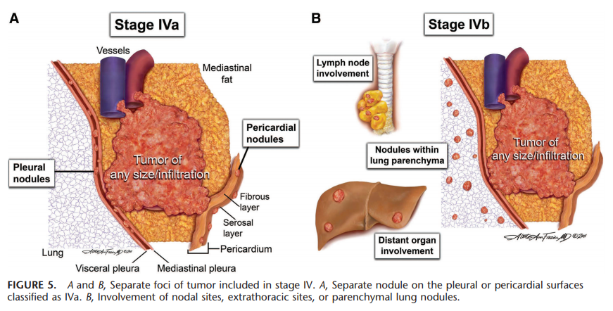
Thymoma and thymic carcinoma staging systems
Masaoka-Koga staging system of thymic tumors:

Source: Jukna A, Jasa M, Mezvevere M, et al. Inside the Mediastinum: the Morphological Spectrum and Stages of Thymic Tumours. Acta Chir Latv. 2016; 16(1):3-8. Available at: https://doi.org/10.1515/chilat-2016-0010.


Source: Detterbeck FC, Nicholson AG, Kondo K, et al. The Masaoka-Koga stage classification for thymic malignancies: clarification and definition of terms. J Thorac Oncol. 2011 Jul;6(7 Suppl 3):S1710-6. Available at: https://doi.org/10.1097/jto.0b013e31821e8cff.
The modified Masaoka staging system separates stage III in IIIa (without invasion of great vessels) and IIIb (with the invasion of great vessels).
Bibliographic reference: NCCN Clinical Practice Guidelines In Oncology (NCCN Guidelines), Thymomas and Thymic Carcinomas, Version 1.2021 - December 4, 2020 [Internet]. Nccn.org. 2020 [cited 26 December 2020]. Available from: https://www.nccn.org/professionals/physician_gls/pdf/thymic.pdf.
The 8th edition thymic TNM stage classifications:


Source: Detterbeck FC. Clinical implication of the new TNM classification of thymic malignancies. J Thorac Dis. 2018 Aug;10(Suppl 22):S2692-S2695. Available at: https://doi.org/10.21037/jtd.2018.08.36.


Source: Detterbeck FC, et al.; Staging and Prognostic Factors Committee; Members of the Advisory Boards; Participating Institutions of the Thymic Domain. The IASLC/ITMIG Thymic Epithelial Tumors Staging Project: proposal for an evidence-based stage classification system for the forthcoming (8th) edition of the TNM classification of malignant tumors. J Thorac Oncol. 2014 Sep;9(9 Suppl 2):S65-72. Available at: https://doi.org/10.1097/jto.0000000000000290.
Relationship between the 8th edition thymic TNM stage and the Masaoka-Kogan system:

Source: Liang G, et al.; Members of the Chinese Alliance for Research in Thymomas. Comparison of the Masaoka-Koga staging and the International Association for the Study of Lung Cancer/the International Thymic Malignancies Interest Group proposal for the TNM staging systems based on the Chinese Alliance for Research in Thymomas retrospective database. J Thorac Dis. 2016 Apr;8(4):727-37. Available at: https://doi.org/10.21037/jtd.2016.03.22.
Monday, 20 July 2020
Tumor budding
Tumor budding is defined as a single tumor cell or a cell cluster of up to 4 tumor cells, it is an independent predictor of lymph node metastasis in pT1 colorectal cancer and an independent predictor of survival in stage II colorectal cancer, and it should be taken into account along with other clinicopathologic factors in a multidisciplinary setting [1].
Tumor budding is counted on hematoxylin-eosin.
Intratumoral tumor budding in colorectal cancer has been shown to be related to lymph node metastasis and it should be included in guidelines/protocols for colorectal cancer reporting [1].
Tumor budding and tumor grade are not the same.
Tumor budding is graded according to its number in a microscopic field with a ×20 objective lens (0.785 mm2) in the hotspot. Tumors with less than five, five to nine, and 10 or more budding foci as classified as grades BD1, BD2, and BD3, respectively [2]. Category BD3 is subclassified as BD3a for tumors with 10 to 19 and BD3b for those with 20 or more budding foci in the hotspot (in a field of 0.785 mm2) at the invasive front [2].
Bibliographic references:
[1] Cho SJ, Kakar S. Tumor Budding in Colorectal Carcinoma: Translating a Morphologic Score Into Clinically Meaningful Results. Arch Pathol Lab Med. 2018;142(8):952-957. Available at: https://doi.org/10.5858/arpa.2018-0082-RA.
[2] Ueno H, et al. Prospective Multicenter Study on the Prognostic and Predictive Impact of Tumor Budding in Stage II Colon Cancer: Results From the SACURA Trial. J Clin Oncol. 2019;37(22):1886-1894. Available at: https://doi.org/10.1200/JCO.18.02059.
Monday, 6 April 2020
Tru-Cut®, core, lance or punch biopsy
It «is when a core of tissue (about 2 millimeters thick) is taken from a lump or tissue using a special needle. Although it can be done in certain circumstances under local anesthetic it is generally performed under a general anesthetic [1].» «The Tru-Cut® biopsy device helps you manually capture high-quality tissue samples with minimal patient trauma [2].» It is a «minimally invasive technique (transcutaneous punctures) for collecting tissue samples by means of special needles that remove a hemicylinder of a lesion, allowing its histological evaluation [3].» «Tru-Cut® biopsy, also called a core, lance, or punch biopsy with a tissue-cutting needle, provides tissue sampling by a percutaneous puncture. These instruments consist of a cutting cannula and a needle with a stylet tip and a semicylinder slot to collect a fresh tissue fragment [4,5]. This technique is normally used in cases when fine-needle aspirates cannot provide a definite diagnosis [5].»
«Tru-Cut® biopsy was first used in 1855 by Duchenne to diagnose Duchenne muscular dystrophy [5].»
«Tru-Cut® biopsy was first used in 1855 by Duchenne to diagnose Duchenne muscular dystrophy [5].»

Tru-Cut® biopsy device
Source: Tru-Cut® Biopsy Device [Internet]. Merit Medical Systems. [cited 2020 Apr 6]. Available from: https://www.merit.com/merit-biopsy/biopsy/soft-tissue-biopsy/tru-cut-biopsy-device/#toggle-id-3.
Tru-Cut® biopsy
Bibliographic references:
[1] Tru-cut biopsy [Internet]. Nottinghamshire Head & Neck Cancer Service. NHS; [cited 2020 Apr 6]. Available from: http://nottshncs.nhs.uk/glossary/tru-cut-biopsy.
[2] Tru-Cut® Biopsy Device [Internet]. Merit Medical Systems. [cited 2020 Apr 6]. Available from: https://www.merit.com/merit-biopsy/biopsy/soft-tissue-biopsy/tru-cut-biopsy-device/#toggle-id-3.
[3] Irion KL, Irion LD, Hochhegger B. (2006). Core Biopsy, Tru Cut Biopsy, Lanceting Puncture or Puncture Biopsy with Tissue Morcelatting Needle? Radiologia Brasileira, 39(4), VII. Available from: https://doi.org/10.1590/S0100-39842006000400003.
[4] Irion KL, Irion LD, Hochhegger B. (2006). Core Biópsia, Tru-Cut biópsia, punção lancetante ou biópsia por punção com agulha fragmentante tecidual (punção fragmentante - PFrag)? Radiologia Brasileira, 39(4), VII. Available from: https://doi.org/10.1590/S0100-39842006000400003.
[5] de Araújo Setin R, Fortes Cirimbelli C, Mazeto Ercolin AC, Pires ST, Disselli T, Ferrarini Nunes Soares Hage MC. Value of artisanal simulators to teach ultrasound-guided percutaneous biopsy using a tru-cut needle for veterinary and medical students. Adv Physiol Educ. 2018;42(2):209–214. Available from: https://doi.org/10.1152/advan.00185.2017.
Wednesday, 29 May 2019
Targeted therapy
There are «some of the differences within cancer cells that enable them to thrive. Targeted therapy refers to treatment with drugs that have been developed to “target” these differences within the cell. Unlike chemotherapy, targeted therapy drugs alter the inner workings of the cell focusing on the part of the cancer cell that makes it different from the normal (...). Because they leave the healthy cells alone the side effects of targeted therapies are different from standard chemotherapy treatments. Targeted therapy works by one of the following: arresting the development of new blood vessels that feed the cancer cell, triggering the immune system to attack the cancer cell, changing proteins within the cancer cell, blocking or turning off signals telling the cancer cell to grow or divide, or carrying toxins directly to the cancer cell [1].»
It is «a type of treatment that uses drugs or other substances to identify and attack specific types of cancer cells with less harm to normal cells. Some targeted therapies block the action of certain enzymes, proteins, or other molecules involved in the growth and spread of cancer cells. Other types of targeted therapies help the immune system kill cancer cells or deliver toxic substances directly to cancer cells and kill them. Targeted therapy may have fewer side effects than other types of cancer treatment. Most targeted therapies are either small molecule drugs or monoclonal antibodies [2].»
«Targeted cancer therapies are drugs or other substances that block the growth and spread of cancer by interfering with specific molecules ("molecular targets") that are involved in the growth, progression, and spread of cancer. (...). Many different targeted therapies have been approved for use in cancer treatment. These therapies include hormone therapies, signal transduction inhibitors, gene expression modulators, apoptosis inducers, angiogenesis inhibitors, immunotherapies, and toxin delivery molecules.
- Hormone therapies slow or stop the growth of hormone-sensitive tumors, which require certain hormones to grow. Hormone therapies act by preventing the body from producing the hormones or by interfering with the action of the hormones. Hormone therapies have been approved for both breast cancer and prostate cancer.
- Signal transduction inhibitors block the activities of molecules that participate in signal transduction, the process by which a cell responds to signals from its environment. During this process, once a cell has received a specific signal, the signal is relayed within the cell through a series of biochemical reactions that ultimately produce the appropriate response(s). In some cancers, the malignant cells are stimulated to divide continuously without being prompted to do so by external growth factors. Signal transduction inhibitors interfere with this inappropriate signaling.
- Gene expression modulators modify the function of proteins that play a role in controlling gene expression.
- Apoptosis inducers cause cancer cells to undergo a process of controlled cell death called apoptosis. Apoptosis is one method the body uses to get rid of unneeded or abnormal cells, but cancer cells have strategies to avoid apoptosis. Apoptosis inducers can get around these strategies to cause the death of cancer cells.
- Angiogenesis inhibitors block the growth of new blood vessels to tumors (a process called tumor angiogenesis). A blood supply is necessary for tumors to grow beyond a certain size because blood provides the oxygen and nutrients that tumors need for continued growth. Treatments that interfere with angiogenesis may block tumor growth. Some targeted therapies that inhibit angiogenesis interfere with the action of vascular endothelial growth factor (VEGF), a substance that stimulates new blood vessel formation. Other angiogenesis inhibitors target other molecules that stimulate new blood vessel growth.
- Immunotherapies trigger the immune system to destroy cancer cells. Some immunotherapies are monoclonal antibodies that recognize specific molecules on the surface of cancer cells. Binding of the monoclonal antibody to the target molecule results in the immune destruction of cells that express that target molecule. Other monoclonal antibodies bind to certain immune cells to help these cells better kill cancer cells.
- Monoclonal antibodies that deliver toxic molecules can cause the death of cancer cells specifically. Once the antibody has bound to its target cell, the toxic molecule that is linked to the antibody — such as a radioactive substance or a poisonous chemical — is taken up by the cell, ultimately killing that cell. The toxin will not affect cells that lack the target for the antibody — i.e., the vast majority of cells in the body.
Bibliographic references:
[1] Cancer-champions.com. (n.d.). Chemotherapy, Immunotherapy, Targeted Therapy. What's the difference? | Cancer-Champions health-care consulting. [online] Available at: https://cancer-champions.com/chemotherapy-immunotherapy-targeted-therapy-whats-the-difference/ [Accessed 29 May 2019].
[2] National Cancer Institute. (n.d.). NCI Dictionary of Cancer Terms. [online] Available at: https://www.cancer.gov/publications/dictionaries/cancer-terms/def/targeted-therapy [Accessed 29 May 2019].
[3] National Cancer Institute. (n.d.). Targeted Cancer Therapies. [online] Available at: https://www.cancer.gov/about-cancer/treatment/types/targeted-therapies/targeted-therapies-fact-sheet [Accessed 29 May 2019].
Sunday, 31 December 2017
TGR (tumor regression grade) in rectal cancer
Pathological features, or TGR, of rectal cancer after preoperative radiochemotherapy:

Source: Dworak O, et al, 1997, cited by Park YJ, Oh BR, Lim SW, Huh JW, Joo JK, Kim YJ, Kim HR. Clinical significance of tumor regression grade in rectal cancer with preoperative chemoradiotherapy. J Korean Soc Coloproctol. 2010 Aug;26(4):279-86. Available at: https://doi.org/10.3393/jksc.2010.26.4.279.
TRG (tumor regression grade) in esophageal carcinoma
Pathological response grading or TRG following neoadjuvant chemoradiation in oesophageal cancer:

Source: Mandard AM, et al., 1994, cited by Gillham CM, Reynolds J, Hollywood D. Predicting the response of localised oesophageal cancer to neo-adjuvant chemoradiation. World J Surg Oncol. 2007 Aug 23;5:97. Available at: https://doi.org/10.1186/1477-7819-5-97.
Wednesday, 10 August 2016
Tpot (potential doubling time without cell loss)
It describes the
proliferation rate of the tissues, which can be measured from
biopsies by flow cytometry after administration in situ to the
patient of a deoxyribonucleic acid thymidine tracer [2]. "This is difficult to determine in vivo." It is "the time which the cell population of tumor doubles if there is no cell loss" [3]. Tp (potential
doubling time or cell doubling time after cell loss) is a little shorter than Tpot (measured
before the tumour has received any cytotoxic treatment) [1].
Pretreatment Tpot measured in human
tumor biopsies [2].
|
||
Tumor
|
Median (days)
|
Range (days)
|
Larynx
|
4
|
2-19
|
Tongue
|
4-6
|
2-16
|
Mouth, cheek
|
3.4
|
2-15
|
Esophagus
|
5
|
2.5-20
|
Cervix
|
5
|
3-20
|
Rectum
|
5
|
3-18
|
Prostate
|
42
|
15-70
|
Breast
|
14
|
3-70
|
Bibliographic references:
[1] Fowler, JF. 21 years of
Biologically Effective Dose. Br J Radiol. 2010 Jul; 83(991): 554–568.
Available at: http://dx.doi.org/10.1259%2Fbjr%2F31372149.
[2] Fowler JF.
The radiobiology of prostate cancer including new aspects of
fractionated radiotherapy. Acta Oncol. 2005;44(3):265-76.
Available at: http://dx.doi.org/10.1080/02841860410002824.
[3] The timely delivery of radical radiotherapy: standards and guidelines for the management of unscheduled treatment interruptions. (2008). 3rd ed. London, United Kingdom: Board of Faculty of Clinical Oncology, The Royal College of Radiologists. Available at: https://www.rcr.ac.uk/sites/default/files/publication/BFCO(08)6_Interruptions.pdf [Accessed 17 Jul. 2016].
[3] The timely delivery of radical radiotherapy: standards and guidelines for the management of unscheduled treatment interruptions. (2008). 3rd ed. London, United Kingdom: Board of Faculty of Clinical Oncology, The Royal College of Radiologists. Available at: https://www.rcr.ac.uk/sites/default/files/publication/BFCO(08)6_Interruptions.pdf [Accessed 17 Jul. 2016].
Sunday, 24 July 2016
Tp (potential doubling time or cell doubling time after cell loss)
Tp is the potential doubling time of tumors [1]. It is the constant repopulation rate, i.e., a constant cell doubling time up to the end of the radiation treatment [2]. It is is the average doubling time of the clonogenic cells in days [3]. For example, in head and neck tumours or cervix cancer, it can be as short as 2 days; therefore one loses up to 1 Gy worth of cell killing when prolonging the course of radiotherapy. Tp it the fastest time in which a tumour can double its volume and depends on cell type and can be of the order of 2 days in fast growing tumours [1]. This "volume doubling time is determined by cell cycle time, growth factor, and rate of cell loss." Tumours such as squamous cel carcinomas have a relatively short volume doubling time; adenocarcinomas have a longer volume doubling time [4]. It can be measured in cell biology experiments (that requires optimal conditions for the tumour and is a worst case scenario) [1].
Patients on treatment should be prioritised according to the volume doubling time. The prolongation of overall treatment time affects treatment outcome or local tumour control in patients with tumours with short Tp [4].
In patients with muscle-invasive transitional cell bladder carcinoma, the clonogen doubling time is about 5–8 days [5].
Patients on treatment should be prioritised according to the volume doubling time. The prolongation of overall treatment time affects treatment outcome or local tumour control in patients with tumours with short Tp [4].
In patients with muscle-invasive transitional cell bladder carcinoma, the clonogen doubling time is about 5–8 days [5].
Bibliographic references:
[1] IAEA Training Material on Radiation Protection in Radiotherapy - Radiation Protection in Radiotherapy - Part 3 - Biological Effects - Lecture 2: High Doses in Radiation Therapy. (2013). [Lectures/Slides]. Vienna, Austria: International Atomic Energy Agency. Available at: https://rpop.iaea.org/RPOP/RPoP/Content/Documents/TrainingRadiotherapy/Lectures/RT03_RadBiol2_RT_WEB.ppt [Accessed 12 Jul. 2016].
[2] Fowler, JF. 21 years of Biologically Effective Dose. Br J Radiol. 2010 Jul; 83(991): 554–568. Availavle at: http://dx.doi.org/10.1259%2Fbjr%2F31372149.
[3] Tomé WA, Fowler JF. On the inclusion of proliferation in tumour control probability calculations for inhomogeneously irradiated tumours. Phys Med Biol. 2003 Sep 21;48(18):N261-8. Available at: http://dx.doi.org/10.1088/0031-9155/48/18/402.
[4] The timely delivery of radical radiotherapy: standards and guidelines for the management of unscheduled treatment interruptions. (2008). 3rd ed. London, United Kingdom: Board of Faculty of Clinical Oncology, The Royal College of Radiologists. Available at: https://www.rcr.ac.uk/sites/default/files/publication/BFCO(08)6_Interruptions.pdf [Accessed 17 Jul. 2016].
[5] Bese NS, Hendry J, Jeremic B. Effects of prolongation of overall treatment time due to unplanned interruptions during radiotherapy of different tumor sites and practical methods for compensation. Int J Radiat Oncol Biol Phys. 2007 Jul 1;68(3):654-61. Available at: http://dx.doi.org/10.1016/j.ijrobp.2007.03.010.
[4] The timely delivery of radical radiotherapy: standards and guidelines for the management of unscheduled treatment interruptions. (2008). 3rd ed. London, United Kingdom: Board of Faculty of Clinical Oncology, The Royal College of Radiologists. Available at: https://www.rcr.ac.uk/sites/default/files/publication/BFCO(08)6_Interruptions.pdf [Accessed 17 Jul. 2016].
[5] Bese NS, Hendry J, Jeremic B. Effects of prolongation of overall treatment time due to unplanned interruptions during radiotherapy of different tumor sites and practical methods for compensation. Int J Radiat Oncol Biol Phys. 2007 Jul 1;68(3):654-61. Available at: http://dx.doi.org/10.1016/j.ijrobp.2007.03.010.
Wednesday, 13 July 2016
Tk (kick-off time or onset time) or Tdelay or Tdel
Tk, Tdelay [1],
or Tdel [2] "represents any delay in the start of tumour cell
repopulation in response to radiation treatment after radiation treatment has
started." Repopulation of tumour cells due to radiation treatment starts
after a passed lag period of Tk treatment days [3]. The repopulation time of tumour cells
appears to vary during radiotherapy; at the commencement it may be slow (e.g., due to hypoxia); however, a certain
time after the first fraction of radiotherapy (kick-off time), repopulation
accelerates [4]. Tk is "the apparent starting time of
rapid compensatory repopulation in tumor or tissue after the start of
treatment, when it is assumed that there are just two rates of cell
proliferation during radiotherapy: zero from start to Tk, then
constant doubling each Tp days [cell doubling time] until
end of treatment at T days [overall treatment time]" [5].
Tk could be between 21 and
32 days in human head and neck tumors [6,7] and non-small-cell lung
cancer, and unlikely may be shorter, but may be longer in prostate cancer [8]. Fowler et al. [9] considered that
prostate cancer would have a TK value
up to 10 times the TK for
head and neck tumors, approximately between 210 and 300 days, and Leborgne et
al. [10] considered a TK for prostate cancer of 52 days. Values for adenocarcinoma of the anus have not been defined [11].
Bibliographic references:
[1] The timely delivery of radical radiotherapy: standards and guidelines for the management of unscheduled treatment interruptions. (2008). 3rd ed. London, United Kingdom: Board of Faculty of Clinical Oncology, The Royal College of Radiologists. Available at: https://www.rcr.ac.uk/sites/default/files/publication/BFCO(08)6_Interruptions.pdf [Accessed 17 Jul. 2016].
[2] Wyatt RM, Beddoe AH, Dale RG. The effects of delays in radiotherapy treatment on tumour control. Phys Med Biol. 2003 Jan 21;48(2):139-55. Available at: http://dx.doi.org/10.1088/0031-9155/48/2/301.
[1] The timely delivery of radical radiotherapy: standards and guidelines for the management of unscheduled treatment interruptions. (2008). 3rd ed. London, United Kingdom: Board of Faculty of Clinical Oncology, The Royal College of Radiologists. Available at: https://www.rcr.ac.uk/sites/default/files/publication/BFCO(08)6_Interruptions.pdf [Accessed 17 Jul. 2016].
[2] Wyatt RM, Beddoe AH, Dale RG. The effects of delays in radiotherapy treatment on tumour control. Phys Med Biol. 2003 Jan 21;48(2):139-55. Available at: http://dx.doi.org/10.1088/0031-9155/48/2/301.
[3] Tomé WA, Fowler JF. On the inclusion of proliferation in tumour control probability calculations for inhomogeneously irradiated tumours. Phys Med Biol. 2003 Sep 21;48(18):N261-8. Available at: http://dx.doi.org/10.1088/0031-9155/48/18/402.
[4] IAEA Training Material on Radiation Protection in Radiotherapy - Radiation Protection in Radiotherapy - Part 3 - Biological Effects - Lecture 2: High Doses in Radiation Therapy. (2013). [Lectures/Slides]. Vienna, Austria: International Atomic Energy Agency. Available at: https://rpop.iaea.org/RPOP/RPoP/Content/Documents/TrainingRadiotherapy/Lectures/RT03_RadBiol2_RT_WEB.ppt [Accessed 12 Jul. 2016].
[5] Fowler, J. (2006). Part I: Basic Concepts in Treatment Planning, 1. Practical Time-Dose Evaluations, or How to Stop Worrying and Learn to Love Linear Quadratics. In: S. Levitt, J. Purdy, C. Perez and S. Vijayakumar, ed., Technical Basis of Radiation Therapy, Practical Clinical Applications, 4th ed. Springer-Verlag Berlin Heidelberg, pp.3-31.
[6] Brenner, DJ. Accelerated repopulation during radiotherapy. Quantitative evidence for delayed onset. Radiat Oncol Invest. 1993;1(3):167–72. Available at: http://dx.doi.org/10.1002/roi.2970010306.
[7] Roberts, SA, Hendry, JH. Time factors in larynx tumor radiotherapy: lag times and intertumor heterogeneity in clinical datasets from four centers. Int J Radiat Oncol Biol Phys. 1999;45(5):1247–57. Available at: http://dx.doi.org/10.1016/s0360-3016(99)00320-x.
[8] Fowler JF. Development of radiobiology for oncology—a personal view. Phys Med Biol. 2006 Jul 7;51(13):R263-86. Available at: http://dx.doi.org/10.1088/0031-9155/51/13/R16.
[9] Fowler, JF, Ritter, MA, Fenwick , JD, Chappell, RJ. How low is the alpha/beta ratio for prostate cancer? In regard to Wang et al., IJROBP 2003;55:194-203, Int J Radiat Oncol Biol Phys. 2003;57(2):593–5. Available at: http://dx.doi.org/10.1016/s0360-3016(03)00364-x.
[8] Fowler JF. Development of radiobiology for oncology—a personal view. Phys Med Biol. 2006 Jul 7;51(13):R263-86. Available at: http://dx.doi.org/10.1088/0031-9155/51/13/R16.
[9] Fowler, JF, Ritter, MA, Fenwick , JD, Chappell, RJ. How low is the alpha/beta ratio for prostate cancer? In regard to Wang et al., IJROBP 2003;55:194-203, Int J Radiat Oncol Biol Phys. 2003;57(2):593–5. Available at: http://dx.doi.org/10.1016/s0360-3016(03)00364-x.
[10] Leborgne, F, Fowler, J, Leborgne, JH, Mezzera, J. Later
outcomes and alpha/beta estimate from hypofractionated conformal
threediomensional radiotherapy versus standard fractionation for localized
prostate cancer. Int J Radiat Oncol Biol Phys. 2012;82(3):1200–7. Available at: http://dx.doi.org/10.1016/j.ijrobp.2010.12.040.
[11] Joon DL, Chao MW, Ngan SY, Joon ML, Guiney MJ. Primary adenocarcinoma of the anus: a retrospective analysis. Int J Radiat Oncol Biol Phys. 1999 Dec 1;45(5):1199-205. Available at: http://dx.doi.org/10.1016/S0360-3016(99)00267-9.
[11] Joon DL, Chao MW, Ngan SY, Joon ML, Guiney MJ. Primary adenocarcinoma of the anus: a retrospective analysis. Int J Radiat Oncol Biol Phys. 1999 Dec 1;45(5):1199-205. Available at: http://dx.doi.org/10.1016/S0360-3016(99)00267-9.
Saturday, 25 June 2016
TARGIT (targeted intraoperative radiotherapy)
It is a technique of giving radiotherapy to the tissues surrounding a cancer after its surgical removal. In patients having
lumpectomy for breast cancer, the TARGIT-A(lone) randomized
controlled trial [1] tested whether
TARGIT within a risk-adapted approach was non-inferior to conventional course
of external beam postoperative radiotherapy given over several weeks. The
conclusion was TARGIT
concurrent with lumpectomy should be considered an option for eligible patients
[2]. The ongoing TARGIT-B(oost) randomized
controlled trial is testing whether TARGIT
tumour bed boost given after lumpectomy for breast cancer in younger patients
or those with a high risk of recurrence is superior to conventional external
beam radiotherapy boost for breast cancer [3].
The TARGIT technique is performed by low
energy X-rays (50kV maximum) at the tip of a tube. The radiation source is
inserted into the tumour bed immediately after excision of the tumour and
switched on for 20-35 minutes to provide intra-operative radiotherapy
accurately targeted to the tissues that are at highest risk of local recurrence
[4].
Other nomenclatures: TARGIT-BQR (boost quality registry) [5], TARGIT-C (consolidation) [5],
TARGIT-D (ductal carcinoma in situ) [6], TARGIT-E (elderly) [7], TARGIT-R (retrospective) [8], TARGIT-US (United States) [9].
Bibliographic references:
[1] Vaidya JS, Wenz
F, Bulsara M, Tobias JS, Joseph
DJ, Keshtgar M, et al; TARGIT trialists'
group. Risk-adapted
targeted intraoperative radiotherapy versus whole-breast radiotherapy for
breast cancer: 5-year results for local control and overall survival from
the TARGIT-A randomised trial. Lancet. 2014 Feb
15;383(9917):603-13. Available at: http://dx.doi.org/10.1016/S0140-6736(13)61950-9.
[2] En.wikibooks.org. (2016). Radiation Oncology/Breast/Partial
breast irradiation - Wikibooks, open books for an open world. [online]
Available at: https://en.wikibooks.org/wiki/Radiation_Oncology/Breast/Partial_breast_irradiation [Accessed 25 Jun. 2016].
[3] Wikipedia. (2016). Targeted
intra-operative radiotherapy. [online] Available at:
https://en.m.wikipedia.org/wiki/Targeted_intra-operative_radiotherapy [Accessed
25 Jun. 2016].
[4]
Ucl.ac.uk. (2016). TARGIT-B.
[online] Available at: http://www.ucl.ac.uk/silva/surgical-interventional-trials-unit/trials/breast/targit-trials/targit-b [Accessed 25 Jun. 2016].
[5] Sperk E, Astor D, Keller A, Welzel G, Gerhardt A, Tuschy B, et al. A cohort analysis to identify eligible patients for intraoperative radiotherapy (IORT) of early breast cancer. Radiat Oncol. 2014; 9: 154. Available at: http://dx.doi.org/10.1186%2F1748-717X-9-154.
[6] Williams, N. and Reynolds, C. (2014). Treating Patients with TARGIT. In: M. Keshtgar, K. Pigott and F. Wenz, ed., Targeted Intraoperative Radiotherapy in Oncology, 1st ed. Springer-Verlag Berlin Heidelberg, pp.141-145.
[7] Neumaier C, Elena S, Grit W, Yasser AM, Uta KT, Anke K, et al. TARGIT-E(lderly)--prospective phase II study of intraoperative radiotherapy (IORT) in elderly patients with small breast cancer. BMC Cancer. 2012 May 8;12:171. Available at: http://dx.doi.org/10.1186/1471-2407-12-171.
[8] Valente
SA, Tendulkar RD, Cherian S, O'Rourke C, Greif JM, Bailey L, et
al. TARGIT-R (Retrospective): North American Experience with Intraoperative
Radiation Using Low-Kilovoltage X-Rays for Breast Cancer. Ann
Surg Oncol. 2016 May 9. Available
at: http://dx.doi.org/10.1245/s10434-016-5240-1.
[9] Clinicaltrials.gov. (2016). Targeted Intraoperative
Radiotherapy United States (TARGIT-US) Registry Trial - Full Text View -
ClinicalTrials.gov. [online] Available at: https://clinicaltrials.gov/ct2/show/NCT01570998 [Accessed 25 Jun. 2016].
Saturday, 21 May 2016
TROG
Trans Tasman Radiation Oncology Group.
Bibliographic reference: Trog.com.au. (2013). TROG Cancer Research - Home. [online] Available at: http://www.trog.com.au/ [Accessed 21 May 2016].
Subscribe to:
Posts (Atom)