Source: Top Papers for 2020 - Radiation Oncology (shared collection) [Internet]. Read.qxmd.com. 2020 [cited 29 December 2020]. Available from: https://read.qxmd.com/collection/22121?sid=adea2753-d897-4e24-bd70-dad41e492317.
Tuesday, 29 December 2020
Saturday, 26 December 2020
Thymoma and thymic carcinoma staging systems
Masaoka-Koga staging system of thymic tumors:

Source: Jukna A, Jasa M, Mezvevere M, et al. Inside the Mediastinum: the Morphological Spectrum and Stages of Thymic Tumours. Acta Chir Latv. 2016; 16(1):3-8. Available at: https://doi.org/10.1515/chilat-2016-0010.


Source: Detterbeck FC, Nicholson AG, Kondo K, et al. The Masaoka-Koga stage classification for thymic malignancies: clarification and definition of terms. J Thorac Oncol. 2011 Jul;6(7 Suppl 3):S1710-6. Available at: https://doi.org/10.1097/jto.0b013e31821e8cff.
The modified Masaoka staging system separates stage III in IIIa (without invasion of great vessels) and IIIb (with the invasion of great vessels).
Bibliographic reference: NCCN Clinical Practice Guidelines In Oncology (NCCN Guidelines), Thymomas and Thymic Carcinomas, Version 1.2021 - December 4, 2020 [Internet]. Nccn.org. 2020 [cited 26 December 2020]. Available from: https://www.nccn.org/professionals/physician_gls/pdf/thymic.pdf.
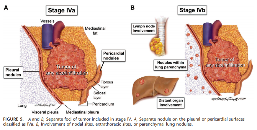
The 8th edition thymic TNM stage classifications:


Source: Detterbeck FC. Clinical implication of the new TNM classification of thymic malignancies. J Thorac Dis. 2018 Aug;10(Suppl 22):S2692-S2695. Available at: https://doi.org/10.21037/jtd.2018.08.36.


Source: Detterbeck FC, et al.; Staging and Prognostic Factors Committee; Members of the Advisory Boards; Participating Institutions of the Thymic Domain. The IASLC/ITMIG Thymic Epithelial Tumors Staging Project: proposal for an evidence-based stage classification system for the forthcoming (8th) edition of the TNM classification of malignant tumors. J Thorac Oncol. 2014 Sep;9(9 Suppl 2):S65-72. Available at: https://doi.org/10.1097/jto.0000000000000290.
Relationship between the 8th edition thymic TNM stage and the Masaoka-Kogan system:

Source: Liang G, et al.; Members of the Chinese Alliance for Research in Thymomas. Comparison of the Masaoka-Koga staging and the International Association for the Study of Lung Cancer/the International Thymic Malignancies Interest Group proposal for the TNM staging systems based on the Chinese Alliance for Research in Thymomas retrospective database. J Thorac Dis. 2016 Apr;8(4):727-37. Available at: https://doi.org/10.21037/jtd.2016.03.22.
Saturday, 12 December 2020
Friday, 11 December 2020
Castration resistant prostate cancer (CRPC)
It is defined as medical or surgical castration, with castrate serum testosterone < 50 ng/dl, or < 1.7 nmol/l, plus one of the following types of progression [1]:
- Biochemical progression:
- Three consecutive rises in total prostate-specific antigen (PSA), at least 1 week apart, resulting in two 50% increases over the nadir, and at least one total PSA > 2 ng/ml [1];
- Or a rising total PSA that is greater than 2 ng/mL higher than the nadir, the rise has to be at least 25% over nadir and the rise has to be confirmed by a second total PSA at least three weeks later [2];
- For CRPC diagnosis, PSA progresses despite secondary hormonal manipulations, including anti‐androgen withdrawal for at least 4 weeks [3].
- Radiologic progression: The appearance of new lesions, either two or more new bone lesions on bone scan or a soft tissue lesion using the Response Evaluation Criteria in Solid Tumours (RECIST).
[1] Cornford P, Bellmunt J, Bolla M, et al. EAU-ESTRO-SIOG Guidelines on Prostate Cancer. Part II: Treatment of Relapsing, Metastatic, and Castration-Resistant Prostate Cancer. Eur Urol. 2017 Apr;71(4):630-642. Available at: https://doi.org/10.1016/j.eururo.2016.08.002.
[2] Scher HI, Halabi S, Tannock I, et al.; Prostate Cancer Clinical Trials Working Group. Design and end points of clinical trials for patients with progressive prostate cancer and castrate levels of testosterone: recommendations of the Prostate Cancer Clinical Trials Working Group. J Clin Oncol. 2008 Mar 1;26(7):1148-59. Available at: https://doi.org/10.1200/jco.2007.12.4487.
[3] Heidenreich A, Aus G, Bolla M, et al.; European Association of Urology. EAU guidelines on prostate cancer. Eur Urol. 2008 Jan;53(1):68-80. Available at: https://doi.org/10.1016/j.eururo.2007.09.002.
Sunday, 15 November 2020
Dx
Dx is defined as the minimum dose to the x% volume of an organ or target receiving the highest dose [1]. Dx is the dose to x% of the organ [2]. Dx is the dose that covers x percent of the structure [3].
Bibliographic references:
[1] Huang EX, Bradley JD, El Naqa I, et al. Modeling the risk of radiation-induced acute esophagitis for combined Washington University and RTOG trial 93-11 lung cancer patients. Int J Radiat Oncol Biol Phys. 2012 Apr 1;82(5):1674-9. Available at: https://doi.org/10.1016/j.ijrobp.2011.02.052.
[2] Chan C, Lang S, Rowbottom C, Guckenberger M, Faivre-Finn C; IASLC Advanced Radiation Technology Committee. Intensity-modulated radiotherapy for lung cancer: current status and future developments. J Thorac Oncol. 2014 Nov;9(11):1598-608. Available at: https://doi.org/10.1097/JTO.0000000000000346.
[3] Livingston GC, Last AJ, Shakespeare TP, et al. Toxicity and dosimetric analysis of non-small cell lung cancer patients undergoing radiotherapy with 4DCT and image-guided intensity modulated radiotherapy: a regional centre's experience. J Med Radiat Sci. 2016 Sep;63(3):170-8. Available at: https://doi.org/10.1002/jmrs.159.
Friday, 30 October 2020
Computed tomography (CT) versus magnetic resonance imaging (MRI) "shades of grey"

Drawing created and kindly provided by @instant.oncology: Wang, L., 2020. Login • Instagram. [online] Instagram.com. Available at: <https://www.instagram.com/p/B-d8KlUAhCP/> [Accessed 30 October 2020].
∝: proportional to; DWI: diffusion-weighted imaging; STIR: short-T1 inversion recovery; Rx: x-rays; Z: atomic number or proton number.
"CT (...) uses kilovoltage x-rays, which interact with matter via photoelectric interactions, and are attenuated depending on the Z-number of the material. (...). Metalwork (such as hip prostheses or dental implants) can also lead to substantial artifacts because they attenuate the x-rays so much that a surrounding “shadow” is created. (...).
MRI (...) does not involve radiation exposure, but (...) [is] also highly prone to artifact by metal implants, even more so than CT. Artifacts tend to affect only CT axial slices containing the implant; but with MRI the distortion can extend to several slices above and below the implant. (...)."
Bibliographic reference: Wang, L., 2020. Login • Instagram. [online] Instagram.com. Available at: <https://www.instagram.com/p/B-d8KlUAhCP/> [Accessed 30 October 2020].
Monday, 12 October 2020
Cutaneous areas at risk for non-melanoma skin cancer
The National Comprehensive Cancer Network (NCCN) guidelines (squamous cell carcinoma [1] and basal cell carcinoma [2]) classify high-risk lesions by size and location, as follows:
- 2 cm or more in diameter in low-risk locations (areas L):
- trunk and extremities, but not including the pretibia, hands, feet, ankles, and nail units.
- 1 cm or more in diameter in moderate-risk locations (area M):
- cheeks, forehead, scalp, neck, and pretibia.
- The area H, independent of size:
- The "mask area" of the face:
- Central face, eyelids, eyebrows, periorbital, nose, lips (cutaneous and vermillion), chin, mandible, preauricular and postauricular skin/sulci, temple, and ear.
- Genitalia, hands, and feet.
Head and neck H and M areas:

Source: [3]

Source: [4]
Body H, M, and L areas [3]:
Bibliographic references:
[1] NCCN Clinical Practice Guidelines in Oncology (NCCN Guidelines), Squamous Cell Skin Cancer, Version 2.2020 - July 14, 2020 [Internet]. Nccn.org. 2020 [cited 12 October 2020]. Available at: https://www.nccn.org/professionals/physician_gls/pdf/squamous.pdf.
[2] NCCN Clinical Practice Guidelines in Oncology (NCCN Guidelines), Basal Cell Skin Cancer, Version 1.2020 - October 24, 2020 [Internet]. Nccn.org. 2020 [cited 12 October 2020]. Available at: https://www.nccn.org/professionals/physician_gls/pdf/nmsc.pdf.
[3] Blechman et al. Application of Mohs micrographic surgery appropriate-use criteria to skin cancers at a university health system. J Am Acad Dermatol. 2014 Jul;71(1):29-35. Available at: https://doi.org/10.1016/j.jaad.2014.02.025.
[4] Dębski T, et al. Basal cell carcinoma. Current views (Part II). Diagnostics and treatment. Borgis - Postępy Nauk Medycznych 2009;9:706-13 [cited 12 October 2020]. Available at: http://ksiaznica.home.pl/pnm/spnm.php?ktory=518.
Sunday, 4 October 2020
Castleman disease
It «is a rare disease of lymph nodes and related tissues. It was first described by Dr. Benjamin Castleman in the 1950s. It is also known as Castleman’s disease, giant lymph node hyperplasia, and angiofollicular lymph node hyperplasia (AFH). Castleman disease is not cancer. Instead, it is called a lymphoproliferative disorder. This means there is an abnormal overgrowth of cells of the lymph system that is similar in many ways to lymphomas (cancers of lymph nodes).
Even though Castleman disease is not officially cancer, one form of this disease (known as multicentric Castleman disease) acts very much like lymphoma. In fact, many people with this disease eventually develop lymphomas. And like lymphoma, Castleman disease is often treated with chemotherapy or radiation therapy.»
«Castleman disease is classified by how much of the body it affects. The main forms of Castleman disease are called localized and multicentric.»
«Infection with certain viruses plays a role in at least some cases of Castleman disease.
Multicentric Castleman disease is more common in people infected with HIV (human immunodeficiency virus), (...). Doctors sometimes group patients with multicentric Castleman disease into those who are infected with HIV (HIV positive) and those who are not infected (HIV negative).
In recent years, it’s become clear that another virus, known as human herpesvirus-8 (HHV-8) or Kaposi sarcoma herpesvirus (KSHV), is often found in the lymph node cells of people with multicentric Castleman disease. In fact, HHV-8 is found in the lymph nodes of nearly all Castleman disease patients who are HIV positive. Some doctors have suggested classifying Castleman disease based on whether the cells contain HHV-8.»
Bibliographic reference: The American Cancer Society medical and editorial content team, 2018. What Is Castleman Disease? [online] Cancer.org. Available at: <https://www.cancer.org/cancer/castleman-disease/about/what-is-castleman-disease.html> [Accessed 4 October 2020].
FoundationOne® Heme test
It «is a comprehensive genomic profiling test combining DNA (deoxyribonucleic acid) sequencing of 406 genes and RNA (ribonucleic acid) sequencing of 265 genes for patients with hematologic malignancies, sarcomas or solid tumors where RNA sequencing is desired [1].» It «detects known, novel, and complex fusion events as well as other common genomic alterations [2]» «(base substitutions, insertion and deletions [indels], [...] copy number alterations [CNAs] [3], and copy number variations [CNVs]) (...) to identify potential targeted therapy options, detect alterations in prognostic genes, and sub-classify sarcoma diagnoses [2].» It identifies «somatic genomic alterations in genes known to be unambiguous drivers of hematologic malignancies (...) and sarcomas [3].» It «is validated to detect the four main classes of genomic alterations in more than 400 cancer-related genes [4].»
Bibliographic references:
[1] Foundation Medicine. 2020. Foundationone Heme. [online] Available at: <https://corpsite.foundationmedicine.com/genomic-testing/foundation-one-heme> [Accessed 4 October 2020].
[2] Foundationmedicine.com. 2020. Foundationone Heme | Foundation Medicine. [online] Available at: <https://www.foundationmedicine.com/test/foundationone-heme> [Accessed 4 October 2020].
[3] Foundationmedicineasia.com. 2017. Foundationone®Heme - Technical Information. [online] Available at: <https://www.foundationmedicineasia.com/content/dam/rfm/apac_v2-en/FoundationOne_Heme_Technical_Information.pdf> [Accessed 4 October 2020].
[4] Foundation Medicine. 2020. Foundation Medicine. [online] Available at: <https://www.foundationmedicine.ca/foundationOneHeme.aspx> [Accessed 4 October 2020].
Monday, 20 July 2020
Tumor budding
Tumor budding is defined as a single tumor cell or a cell cluster of up to 4 tumor cells, it is an independent predictor of lymph node metastasis in pT1 colorectal cancer and an independent predictor of survival in stage II colorectal cancer, and it should be taken into account along with other clinicopathologic factors in a multidisciplinary setting [1].
Tumor budding is counted on hematoxylin-eosin.
Intratumoral tumor budding in colorectal cancer has been shown to be related to lymph node metastasis and it should be included in guidelines/protocols for colorectal cancer reporting [1].
Tumor budding and tumor grade are not the same.
Tumor budding is graded according to its number in a microscopic field with a ×20 objective lens (0.785 mm2) in the hotspot. Tumors with less than five, five to nine, and 10 or more budding foci as classified as grades BD1, BD2, and BD3, respectively [2]. Category BD3 is subclassified as BD3a for tumors with 10 to 19 and BD3b for those with 20 or more budding foci in the hotspot (in a field of 0.785 mm2) at the invasive front [2].
Bibliographic references:
[1] Cho SJ, Kakar S. Tumor Budding in Colorectal Carcinoma: Translating a Morphologic Score Into Clinically Meaningful Results. Arch Pathol Lab Med. 2018;142(8):952-957. Available at: https://doi.org/10.5858/arpa.2018-0082-RA.
[2] Ueno H, et al. Prospective Multicenter Study on the Prognostic and Predictive Impact of Tumor Budding in Stage II Colon Cancer: Results From the SACURA Trial. J Clin Oncol. 2019;37(22):1886-1894. Available at: https://doi.org/10.1200/JCO.18.02059.
Haggitt classification of colorectal polyps

Source: Bujanda L, et al. Malignant colorectal polyps. World J Gastroenterol. 2010;16(25):3103-3111. Available at: https://doi.org/10.3748/wjg.v16.i25.3103.
Narrow‐band imaging (NBI) International Colorectal Endoscopic (NICE) classification of colorectal tumors

WHO: World Health Organization.
Source: Sano Y, et al. Narrow-band imaging (NBI) magnifying endoscopic classification of colorectal tumors proposed by the Japan NBI Expert Team. Dig Endosc. 2016;28(5):526-533. Available at: https://doi.org/10.1111/den.12644.
Wednesday, 8 July 2020
Liver segments
Couinaud classification of liver anatomy:

Source: Chen Y, et al. Functional Region Annotation of Liver CT Image Based on Vascular Tree. Biomed Res Int. 2016;2016:5428737. Available at: https://doi.org/10.1155/2016/5428737.
Sunday, 5 July 2020
Saturday, 4 July 2020
Sunday, 31 May 2020
Imagining image guidance in liver SBRT
November 7-9, 2013, Dr. Laura Dawson, MD, The Princess Margaret Cancer Centre, Toronto, Canada.
SBRT for liver tumors
Feb 16, 2010: Bryan W. Chang, M.D., Yale Cancer Center, New Haven, Connecticut, United States.
Tuesday, 26 May 2020
Pneumonitis of radiation
Grading systems for evaluation of radiation pneumonitis:
 |
| Source: Jain V, Berman AT. Radiation Pneumonitis: Old Problem, New Tricks. Cancers (Basel). 2018;10(7):222. Published 2018 Jul 3. Available at: https://doi.org/10.3390/cancers10070222. |
Treatment: «Corticosteroids are commonly used for the treatment of radiation pneumonitis. Prednisone is started at 1 mg/kg/day upon diagnosis and is continued for several weeks and then tapered slowly. Other agents that have been used for the treatment of radiation pneumonitis include azathioprine and cyclosporin A.» Source: Virna Ignacio Almuete. Treatment for Radiation Pneumonitis - Medscape - Jan 06, 2003. [online] Available at: <https://www.medscape.com/viewarticle/446886> [Accessed 26 May 2020].
Monday, 25 May 2020
Sunday, 17 May 2020
ARC (The Anatomy and Radiology Contouring) Bootcamp
Source: ARC Bootcamp. n.d. Anatomy And Radiology Contouring Bootcamp For Radiation Oncology Residents. [online] Available at: <https://www.arcbootcamp.com/> [Accessed 16 May 2020].
Monday, 11 May 2020
Survey - whole pelvic radiation in prostate cancer - yes or no?
Click here to start (< 5 min): https://docs.google.com/forms/d/e/1FAIpQLSfyBE0ADMlBdvatmvT1XbpiHdLRiALFtm-hTQLNxWE4hJOw7Q/viewform
Survey about prostate cancer radiotherapy to find if Radiation Oncologists:
- used to irradiate the whole pelvis or do not;
- which doses use to give;
- which fractionation use to give.
The purpose of this survey is to learn about the experiences of different Departments around the world and publish the results.
This survey is voluntary and confidential.
Thank you for your time!
Tuesday, 28 April 2020
SPO (Sociedade Portuguesa de Oncologia)
SPO (Sociedade Portuguesa de Oncologia [Portuguese Society of Oncology]).
Reference: SPO. n.d. Sociedade Portuguesa Oncologia. [online] Available at: <https://www.sponcologia.pt/pt/> [Accessed 28 April 2020].
Survey - conhecimento e percepções sobre o Registo Oncológico Nacional (Portuguese) (knowledge and perceptions about the National Cancer Registry)
Survey on knowledge and perceptions about the Portuguese RON (Registo Oncológico Nacional [National Cancer Registry]): https://forms.gle/JqyQRQZho2q7KcvF8
This questionnaire was prepared by the Oncology Data Working Group of the Portuguese Society of Oncology (SPO).
Sunday, 26 April 2020
SEOR
Sociedad Española de Oncología Radioterápica (SEOR) (Spanish Society of Radiation Oncology).
Bibliographic reference: SEOR. n.d. Inicio - SEOR. [online] Available at: <http://www.seor.es/> [Accessed 26 April 2020].
COVID-19 RadOnc Forum
[Spanish]
COVID-19 RadOnc Forum: una plataforma para evaluar diariamente el impacto de la pandemia COVID en la Oncología Radioterápica española (a platform to daily assess the impact of the COVID pandemic on Spanish Radiation Oncology).
More information on SEOR.
Wednesday, 22 April 2020
Online certificate, diploma, M.Sc in advanced radiotherapy practice
Deadline: July 30th, 2020
Source: Tcd.ie. n.d. Online Taught Postgraduate Education - School Of Medicine - Trinity College Dublin. [online] Available at: <https://www.tcd.ie/medicine/radiation-therapy/postgraduate/ARP-online.php> [Accessed 21 April 2020].
Monday, 20 April 2020
IASLC (International Association for the Study of Lung Cancer)
Bibliographic reference: International Association for the Study of Lung Cancer | IASLC [Internet]. Iaslc.org. [cited 20 April 2020]. Available from: https://www.iaslc.org/.
Monday, 6 April 2020
Tru-Cut®, core, lance or punch biopsy
It «is when a core of tissue (about 2 millimeters thick) is taken from a lump or tissue using a special needle. Although it can be done in certain circumstances under local anesthetic it is generally performed under a general anesthetic [1].» «The Tru-Cut® biopsy device helps you manually capture high-quality tissue samples with minimal patient trauma [2].» It is a «minimally invasive technique (transcutaneous punctures) for collecting tissue samples by means of special needles that remove a hemicylinder of a lesion, allowing its histological evaluation [3].» «Tru-Cut® biopsy, also called a core, lance, or punch biopsy with a tissue-cutting needle, provides tissue sampling by a percutaneous puncture. These instruments consist of a cutting cannula and a needle with a stylet tip and a semicylinder slot to collect a fresh tissue fragment [4,5]. This technique is normally used in cases when fine-needle aspirates cannot provide a definite diagnosis [5].»
«Tru-Cut® biopsy was first used in 1855 by Duchenne to diagnose Duchenne muscular dystrophy [5].»
«Tru-Cut® biopsy was first used in 1855 by Duchenne to diagnose Duchenne muscular dystrophy [5].»

Tru-Cut® biopsy device
Source: Tru-Cut® Biopsy Device [Internet]. Merit Medical Systems. [cited 2020 Apr 6]. Available from: https://www.merit.com/merit-biopsy/biopsy/soft-tissue-biopsy/tru-cut-biopsy-device/#toggle-id-3.
Tru-Cut® biopsy
Bibliographic references:
[1] Tru-cut biopsy [Internet]. Nottinghamshire Head & Neck Cancer Service. NHS; [cited 2020 Apr 6]. Available from: http://nottshncs.nhs.uk/glossary/tru-cut-biopsy.
[2] Tru-Cut® Biopsy Device [Internet]. Merit Medical Systems. [cited 2020 Apr 6]. Available from: https://www.merit.com/merit-biopsy/biopsy/soft-tissue-biopsy/tru-cut-biopsy-device/#toggle-id-3.
[3] Irion KL, Irion LD, Hochhegger B. (2006). Core Biopsy, Tru Cut Biopsy, Lanceting Puncture or Puncture Biopsy with Tissue Morcelatting Needle? Radiologia Brasileira, 39(4), VII. Available from: https://doi.org/10.1590/S0100-39842006000400003.
[4] Irion KL, Irion LD, Hochhegger B. (2006). Core Biópsia, Tru-Cut biópsia, punção lancetante ou biópsia por punção com agulha fragmentante tecidual (punção fragmentante - PFrag)? Radiologia Brasileira, 39(4), VII. Available from: https://doi.org/10.1590/S0100-39842006000400003.
[5] de Araújo Setin R, Fortes Cirimbelli C, Mazeto Ercolin AC, Pires ST, Disselli T, Ferrarini Nunes Soares Hage MC. Value of artisanal simulators to teach ultrasound-guided percutaneous biopsy using a tru-cut needle for veterinary and medical students. Adv Physiol Educ. 2018;42(2):209–214. Available from: https://doi.org/10.1152/advan.00185.2017.
Thursday, 19 March 2020

Sunday, 15 March 2020
Recommendations of the Brazilian Radiotherapy Society on COVID-19
Recommendations of the SBRT (Sociedade Brasileira de Radioterapia [Brazilian Radiotherapy Society]) on COVID-19 (Coronavirus disease 2019):
[Portuguese]
Link to clarifications from the SBRT on the care to be taken with patients undergoing radiation therapy during the COVID-19 pandemic: http://sbradioterapia.com.br/noticias/radioterapia-e-ncovid-19-esclarecimentos-da-sbrt/ [Portuguese]. Accessed March 16, 2020.
Friday, 31 January 2020
2020 ESMO awards

Deadline for nominations: 16 February 2020.
Nominate here: 2020 ESMO Award Nominations. ESMO. https://www.esmo.org/career-development/awards/esmo-award-nominations?utm_campaign=2020%20ESMO%20Awards&utm_source=hs_email&utm_medium=email&utm_content=82702576&_hsenc=p2ANqtz-917J9qQf3PoiJdQ31axYuQiW6rm8pkQgp8wDmxQ5CsS9r-duoYXvYjsSYx-6IijSkwgNUEjU3lvoGSgmOre7nynDFv9g&_hsmi=82702576. Published 2020. Accessed January 31, 2020.
Monday, 6 January 2020
Rs of radiobiology
The most important biological factors influencing tumor and normal tissue responses to fractionated treatment are referred to as the Rs [1]:
- Repair [1]/Recovery [2];
- Re-assortment/Redistribution [1,2];
- Repopulation [1,2];
- Reoxygenation [1,2];
- Radiosensitivity [1,2];
- Reactivation of anti-tumor immune response [1];
- intrinsic Radiosensitivity [2];
- iRradiated volume [2];
- long-term Restoration [2];
- molecular Radiopathology [2].
Bibliographic references:
[1] Boustani J, Grapin M, Laurent PA, Apetoh L, Mirjolet C. The 6th R of Radiobiology: Reactivation of Anti-Tumor Immune Response. Cancers (Basel). 2019 Jun 20;11(6):860. Available at: https://dx.doi.org/10.3390%2Fcancers11060860.
[2] Dörr W. Radiobiology of tissue reactions. Ann ICRP. 2015;44(1 Suppl):58–68. Available at: https://doi.org/10.1177/0146645314560686.
Wednesday, 1 January 2020
PD-1 checkpoint inhibitors
A PD-1 (programmed cell death protein 1) inhibitor fairy tale:
How do tumors use PD-1 to cause T-cell exhaustion? Learn how overexpression of PD-1 receptors on T cells may lead to T-cell exhaustion:
ESTRO-ASTRO definition of oligometastatic disease - public comment survey
The deadline to submit comments is Monday, January 6th, 2020: ESTRO-ASTRO definition of oligometastatic disease - public comment survey.
Subscribe to:
Posts (Atom)